WHEN YOUR AMH VALUE IS ABOVE 3.5NG/ML AND YOU DON’T HAVE SIGNS AND SYMPTOM OF PCOS.
WHEN YOUR AMH VALUE IS ABOVE 3.5NG/ML AND YOU DON’T HAVE SIGNS AND SYMPTOM OF PCOS. Anti-Müllerian Hormone (AMH) is a crucial marker used to assess a woman’s ovarian reserve in fertility evaluations. The typical reference range for AMH is approximately 1.0 to 3.5 ng/mL. Values below 1.0 ng/mL may indicate diminished ovarian reserve, while levels above 3.5 ng/mL can be suggestive of Polycystic Ovary Syndrome (PCOS). However, it’s important to note that not all women with elevated AMH levels present with the classical symptoms of PCOS, such as irregular menstrual cycles, acne, or anovulation. This condition is often referred to as “subclinical” or “silent PCOS”, where hormonal imbalance exists without obvious external signs. A high AMH level without symptoms may still affect egg quality and fertilization potential, potentially impacting conception outcomes. It is essential not to rely solely on regular menstruation or ovulation as indicators of reproductive health. If your AMH is above 3.5 ng/mL, or falls below 1.0 ng/mL, it is advisable to consult a fertility specialist for comprehensive evaluation and guidance on appropriate treatment or management strategies.
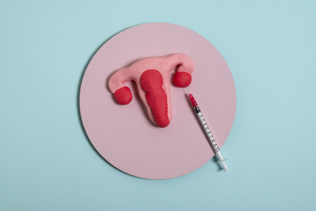
DIFFERENT TYPES OF FALLOPIAN TUBES DAMAGE.
DIFFERENT TYPES OF FALLOPIAN TUBES DAMAGE. Fallopian tube damage is a significant factor in female infertility. Common types of tubal pathology include: 1. Tubal Blockage: Where the fallopian tube is not patent (i.e., not open), preventing the passage of sperm or egg. 2. Hydrosalpinx: A condition where the tube becomes swollen and fluid-filled due to chronic inflammation, often impairing fertility. 3. Fimbrial Adhesions: Adhesions or scarring at the fimbrial end of the tube (the finger-like projections that help capture the egg from the ovary), which can block egg pick-up and transport. If one fallopian tube is damaged or surgically removed commonly after an ectopic pregnancy the other may still function and allow for natural conception. However, if one tube has hydrosalpinx, the toxic fluid it contains may backflow into the uterus and impair implantation, thereby affecting both natural and assisted conception (including IVF). A potential intervention is hydrotubation or tubal flushing, which aims to restore tubal patency. However, this procedure carries no guarantee of success and outcomes remain uncertain. After three months of any tubal treatment, a follow-up Hysterosalpingography (HSG) is recommended to reassess tubal status. If the tubes remain blocked or hydrosalpinx persists, in vitro fertilization (IVF) may be the most appropriate option for achieving pregnancy.
WHEN THERE IS NO GOOD RESULT FROM THE TREATMENT OF SEMEN PROBLEM.
WHEN THERE IS NO GOOD RESULT FROM THE TREATMENT OF SEMEN PROBLEM. There are instances where men undergo treatment for abnormal semen parameters, yet see little or no improvement in their semen analysis. For example, the sperm count may remain persistently low between 2 million and 10 million per milliliter or may even decline further, despite ongoing medication or therapy. In some cases, hormonal profiles and scrotal ultrasound results appear normal, yet sperm count, motility, and morphology fail to improve. One often overlooked cause is a blockage or dysfunction in the epididymis, the coiled tube located at the back of the testicle where sperm is stored and matures. If this duct is obstructed or not functioning properly, sperm cannot pass through effectively, and medical treatments aimed at boosting sperm production may not yield results. In such cases, it is essential to consult a qualified fertility specialist or andrologist who can identify the underlying issue and recommend appropriate management options, which may include further diagnostic evaluation, surgical intervention, or assisted reproductive techniques such as IVF with ICSI.
SEVERE LOW SPERM COUNT AND AZOOSPERMIA
SEVERE LOW SPERM COUNT AND AZOOSPERMIA If you have been diagnosed with azoospermia (complete absence of sperm in the ejaculate) or severe oligospermia (sperm count less than 5 million/ml), it’s important to understand that the chances of spontaneous recovery with medication alone are often limited. Be cautious of anyone claiming to have a guaranteed cure. There are two main types of azoospermia: 1. Obstructive Azoospermia This occurs when sperm production in the testicles is normal, but a blockage prevents the sperm from reaching the ejaculate. Common causes include obstructions in the epididymis, vas deferens, or ejaculatory ducts, and may present as an epididymal cyst. These cases are often challenging and require evaluation by a urologist or male fertility specialist for possible surgical correction or assisted reproductive options like sperm retrieval. 2. Non-Obstructive Azoospermia This form results from impaired or absent sperm production by the testicles. In such cases, a scrotal ultrasound and hormonal profile (e.g., FSH, LH, testosterone) are essential to assess testicular function. Possible causes include undescended testes (cryptorchidism) or testicular failure due to heat damage or developmental issues. If the testes did not properly descend during childhood and remained inside the body beyond puberty, heat exposure may have irreversibly damaged sperm-producing cells. A quick self-check: normally, both testicles should be present in the scrotal sac, located below the penis. During cold temperatures, the scrotum may contract, but the testes should not retract entirely into the body. If you suspect you have a sperm production issue or have been diagnosed with azoospermia or severe oligospermia, consult a fertility scientist.